High-Density CMOS-MEA Reveals Single-Cell Activity and Drug Responses in DRG Neurons
# Unraveling Neural Activity: High-Resolution Insights into Pain and Drug Effects
This study examines the spontaneous activity of dorsal root ganglion (DRG) neurons and their response to pain-related compounds at a single-cell level using high-density CMOS-MEA technology. DRG neurons, known for their diverse soma sizes, were cultured for six weeks in vitro (WIV) and recorded with high spatial resolution.
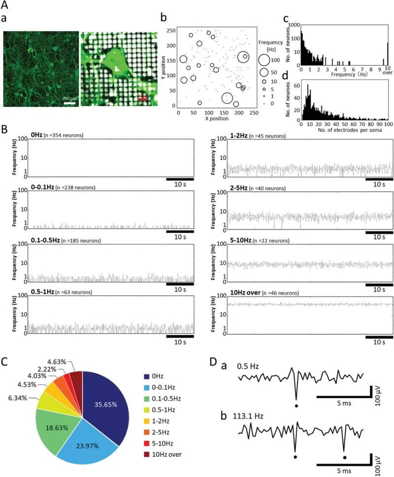
Spontaneous activity was detected in 993 neurons across seven wells, with each soma covering an average of 23.68 electrodes. Unlike central nervous system neurons, DRG neurons lacked synchronized firing and exhibited individual spontaneous activity patterns. The firing frequencies ranged from 0 Hz to over 100 Hz, with the majority of neurons showing low-frequency activity. Notably, 4.63% of neurons exhibited high-frequency firing above 100 Hz, with a peak rate of 113.1 Hz.
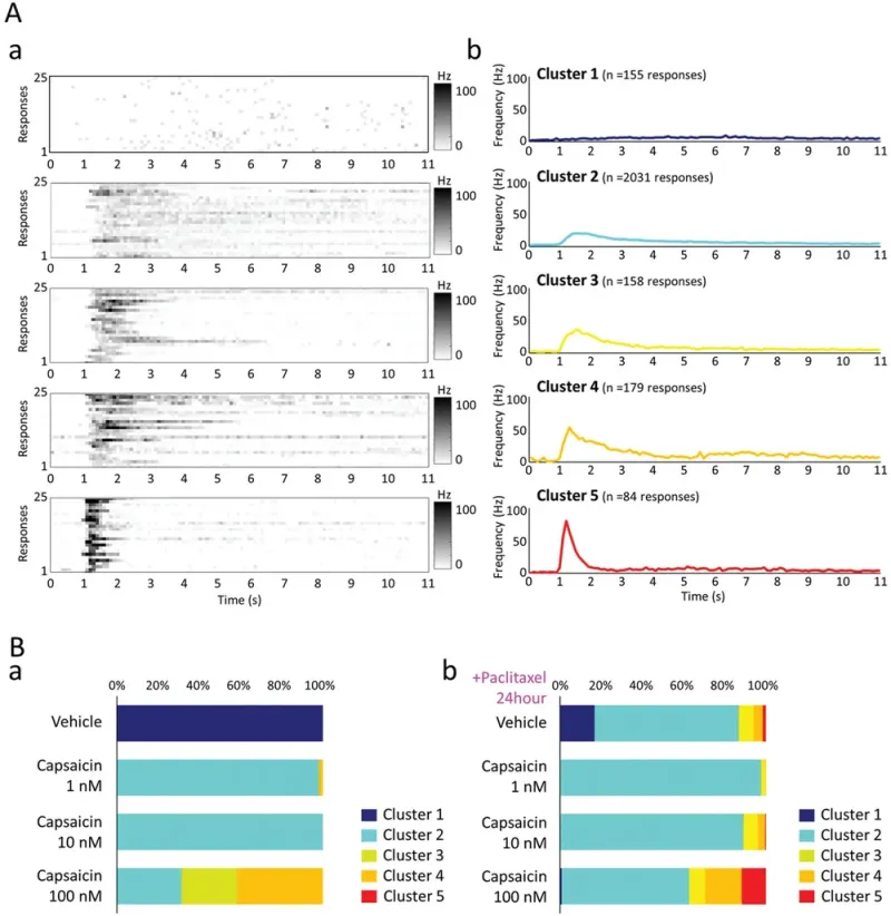
Further analysis assessed the response of DRG neurons to capsaicin, a TRPV1 channel agonist, and the effects of paclitaxel, an anticancer drug. Neurons exhibited five distinct capsaicin-evoked response patterns, ranging from weak activation to strong peak firing (up to 80.2 Hz). Capsaicin-induced responses were concentration-dependent, with higher doses increasing the proportion of highly reactive neurons. Pre-exposure to paclitaxel altered response distributions, with stronger activation of the TRPV1 channel observed at higher capsaicin concentrations.
This study highlights the capability of HD-CMOS-MEA to analyze spontaneous and evoked neuronal activity at single-cell resolution. The findings suggest that DRG neuron activity patterns can be used to evaluate pain responses and drug effects, providing valuable insights into neuropathic pain mechanisms and potential therapeutic interventions.

Spontaneous activity frequency distribution in cultured DRG neurons. A) Immunostaining image of DRG neurons (β-tubuline III) at 6 WIV (weeks in vitro) cultured on HD-CMOS-MEA (a). Enlarged view near the soma (b). DRG neurons detected on the 235 (X-axis) × 252 (Y-axis) = 59220 electrodes and spontaneous activity frequency. The size of the circle indicates the frequency of spontaneous activity from 0 Hz to 100 Hz (c). Histogram of spontaneous activity frequencies of DRG neurons (bin = 100 ms, n = 993 neurons, n = 7 HD-CMOS-MEAs) (d). Histogram of number of electrodes per soma calculated from spontaneous activity of DRG neurons (bin = 1 electrodes, n = 993 neurons, n = 7 HD-CMOS-MEAs). B) The spontaneous activity of single DRG neurons is a typical firing pattern per minute in each frequency band. C) Percentage of DRG neurons in each spontaneous firing frequency band. D) Comparison of voltage waveforms of DRG neurons that exhibited low-frequency firing (a) and exhibited firing rates of >100 Hz (b). Dots show spikes.

Responses to capsaicin in the presence and absence of paclitaxel. A) Clustering of evoked responses in capsaicin administration. Induced response of 25 typical neurons in each of the 5 clusters (bin = 100 ms). The firing frequency of 0 to 100 Hz is shown in gray scale (a). Time course of average firing frequency for each of the 5 clusters (b). B) Cluster ratio of the evoked response to capsaicin 1, 10, 100 nM administration (a). Cluster ratio of evoked response to capsaicin 1, 10, 100 nM administration in the presence of paclitaxel 10 nM for 24 h (b).